Sleepy Eye Medical Center offers a wide range of imaging services that are available upon provider referral. Learn helpful information about these services, how to prepare and what to expect by clicking below.
For more information about our imaging services, call 507-794-8469.
CT
Computer Tomography (CT)
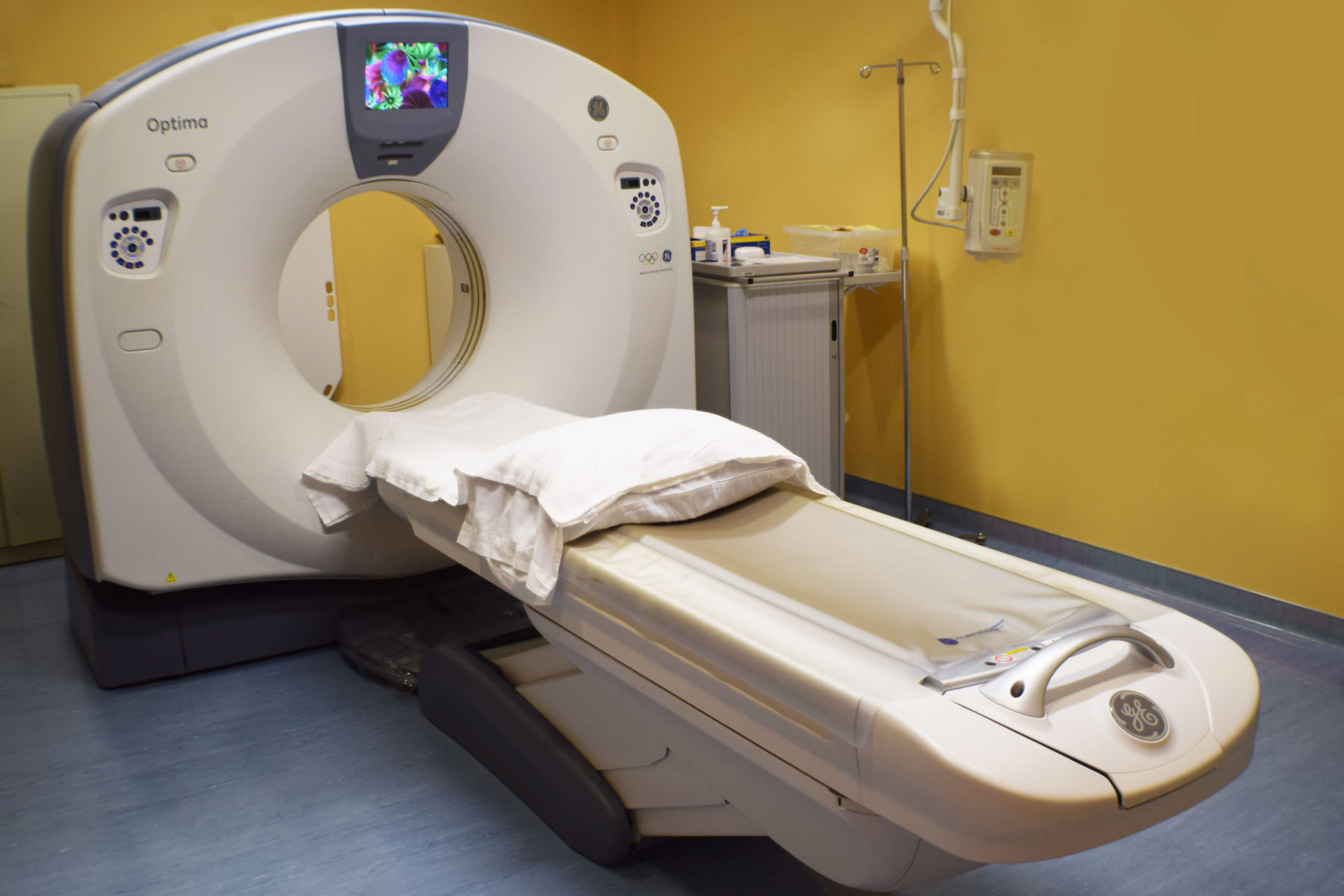
 A Computer Tomography scan (CT) is an imaging test used to take pictures of parts of the body to create detailed images of internal bones, blood vessels and organs.
- How to Prepare
Avoid wearing jewelry or clothing with metal on it, especially if that area is being scanned. In some instances, your provider may order an intravenous (IV) contrast dye injection or oral contrast liquid to drink. This will require you to refrain from eating or drinking three hours prior. Tests using oral constrasts take longer, lasting approximately 1 ½ hours. Please notify an imaging technologist if there’s a chance you could be pregnant. - What to Expect
During the scan, you will lie flat on an imaging table that moves through a doughnut-shaped tube. If contrast fluid is required, you may feel a warm sensation throughout your body as the fluid travels. Scans typically last 10-30 minutes. - Getting Your Results
A radiologist will interpret the images, formulate a report and send the results to your provider. Your provider will contact you to discuss your results.
Lung Cancer Screenings
Lung Cancer Screenings (CT)
Sleepy Eye Medical Center offers lung cancer screening counseling and shared decision-making visits for smokers. After a complete assessment with your provider, you may qualify for annual lung cancer screenings using low dose computed tomography (LDCT).
Lung cancer screening is a preventative service offered to individuals who meet specific criteria, particularly those with a history of smoking. Eligibility includes:
-
Ages 50–77 for Medicare beneficiaries
-
Ages 50–80 for most other insurance plans
-
A smoking history of at least one pack per day for 20 years
-
Currently smoking or having quit within the past 15 years
-
No signs or symptoms of lung cancer
A written order from a healthcare provider is required to schedule a screening at Sleepy Eye Medical Center. Please contact your insurance provider for details on coverage.
- How to Prepare
Make an appointment to complete an assessment with your provider in order to determine if you qualify for annual lung cancer screenings. Preliminary assessments may be completed online at shouldiscreen.com. Avoid wearing jewelry or clothing with metal on it, especially if that area is being scanned. - What to Expect
During the scan, you will lie flat on an imaging table that moves through a doughnut-shaped tube. Contrast solution is not required for this exam. Scans last approximately 10 minutes. - Getting Your Results
A radiologist will read and analyze the images, formulate a report and send the results to your provider. Your provider will mail you a letter containing your results.
DXA
Bone Densitometry (DXA)
Bone density scanning is an enhanced form of Dual-energy X-ray Absorptiometry (DXA) that is used to measure bone loss. Bone density scans safely, accurately and painlessly measure bone density.
The chance of bone fractures and risk of developing osteoporosis increase as a person ages. A DXA scan can help detect bone conditions early and prevent them from getting worse.
- How to Prepare
Refrain from taking calcium supplements 24 hours prior to your exam. If you’ve recently had an exam that required a contrast solution, it is recommended to wait one week before having a DXA scan.
Please notify an imaging technologist if there’s a chance you could be pregnant.
- What to Expect
During the exam, you will lay on a padded table while a mechanical arm passes over two or more areas of the body, typically the hips and spine. The procedure takes approximately 20 minutes to complete. - Getting Your Results
A radiologist will read and analyze the images, formulate a report and send the results to your provider. Your provider will mail you a letter containing your results.
MRI
Magnetic Resonance Imaging (MRI) – mobile truck
Magnetic resonance imaging (MRI) uses a magnetic field, radio waves and computer to take detailed pictures of the spine, joints and organs that cannot be seen with other imaging devices.
- How to Prepare
Dress comfortable and avoid wearing clothing or jewelry that contain metal. You will not be able to have an MRI if you have a pacemaker. If you have questions or a fear of being enclosed in small or narrow spaces (claustrophobia), share these concerns with your provider prior to your scan. - What to Expect
During an MRI, you will lie on a table that slowly moves toward the center of the scanner. It is important to remain as still as possible during the procedure. On occasion, some patients are given an IV contrast solution to improve the image quality. While the scan is painless, you will likely hear various clicking, tapping and buzzing noises – this is normal. Ear plugs will be provided to lessen the intensity of these sounds. The duration of your scan depends on the area(s) of your body being scanned. Should you have any questions or concerns during the process, you will be able to communicate them to the imaging staff. - Getting Your Results
A radiologist will interpret the images, formulate a report and send the results to your provider. Your provider will contact you to discuss your results.
Mammograms
Mammograms
 A mammogram is an X-Ray of the breast. Sleepy Eye Medical Center offers two-dimensional (2-D) and three-dimensional (3-D) mammography to screen for breast cancer and evaluate other breast abnormalities. Mammograms are the best way to find breast cancer early, when it is easier to treat and before it is big enough to feel or cause symptoms. In addition, SEMC offers limited diagnostic breast imaging.
Early detection plays an important role in the successful treatment of breast cancer. For this reason, SEMC providers recommend annual mammograms for women beginning at age 40.
What is 3-D mammography? How is it different than 2-D mammography?
Three-dimensional mammography, also known as breast tomosynthesis, creates three-dimensional images of your breast with clarity and detail by imaging the breast in multiple layers. By viewing each layer separately, a potential cancer is less likely to be obstructed by overlapping breast tissue. In turn, fewer biopsies and additional tests are needed.
Two-dimensional mammography is a standard mammogram that creates a flat image of the breast. They provide safe, reliable results but breast tissue can overlap during the imaging process and lead to inconclusive results. In turn, additional testing may be needed.
We encourage you to discuss these screening options with your provider and contact your insurance company to see what is and isn't covered under your plan.
- How to Prepare
Dress comfortable and avoid wearing deodorant before your mammogram. While some women find mammograms to be uncomfortable, others find them painful but tolerable. Consider taking an over-the-counter pain medication (aspirin, acetaminophen or ibuprofen) about an hour before your exam to ease discomfort. If possible, schedule your mammogram for a time when your breasts aren’t likely to be tender. For women who haven’t gone through menopause, it is recommended to have an exam the week after your menstrual period. - What to Expect
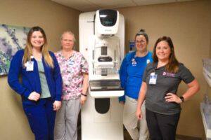
When you arrive, you will be given a gown and asked to remove any jewelry and clothing from the waist up. During the exam, you will be asked to stand in front of the mammography machine and rest one of your breasts on a platform. The mammography technician will raise and lower this platform to match your height. A plate will then gradually apply pressure to your breast, spreading the tissue so that any small abnormalities can be seen. Please stand as still as possible during the imaging process. This procedure will be repeated for your second breast. Four images will be taken: one from the top and one from the side of each breast. The exam takes approximately 15 minutes. - Getting Your Results
A radiologist will interpret the images, formulate a report and send the results to your provider. Your provider will mail you a letter containing your results.
Nuclear Medicine
Nuclear Medicine – mobile truck
Nuclear medicine imaging procedures are used to diagnose certain illnesses using a radioactive substance (tracer). This substance is injected or swallowed, depending on the type of exam. A special camera that detects radiation is placed over your body to take images. These images appear on a computer and show where in the body the radioactive substance accumulates.
- How to Prepare
Some exams require little to no preparation, whereas others require fasting or no consumption of caffeine prior. A member of our staff will provide instructions specific for your procedure when you schedule your exam. - What to Expect
During the exam, you will be given a small dosage of a radioactive substance intravenously (IV) or orally. The scan itself takes approximately 30-60 minutes. Depending on the type of scan being performed, additional time may be required. - Getting Your Results
A radiologist will interpret the images, formulate a report and send the results to your provider. Your provider will contact you to discuss your results.
Ultrasound
Ultrasound (mobile)
 Ultrasound, or sonography, uses high-frequency sound waves to create pictures of structures within the body. Some ultrasound exams are performed outside of body using a transducer (hand-held device), while others involve the placement of the device inside the body.
Sleepy Eye Medical Center provides various ultrasound procedures, including abdominal, pregnancy and vascular ultrasounds.
Abdominal Ultrasounds
An abdominal ultrasound is performed to identify abnormalities of the gallbladder, kidneys, liver, pancreas, spleen or aorta (the large blood vessel that comes from the heart).
- How to Prepare
Before your exam, refrain from drinking, eating, smoking or chewing gum 8 hours prior. - What to Expect
During the exam, you will lie on an examination table and a licensed sonographer will apply gel to your abdomen. He or she will move a hand-held device over your skin. Images will show up on a computer screen and be recorded, along with measurements. The procedure takes approximately 30-45 minutes. - Getting Your Results
A radiologist will interpret the images, formulate a report and send the results to your provider. Your provider will contact you to discuss your results.
Pregnancy Ultrasounds
A pregnancy ultrasound is performed to produce images of your baby in the uterus. Fetal ultrasounds evaluate your baby’s growth and development.
An initial fetal ultrasound may be performed during the first trimester of pregnancy. In uncomplicated pregnancies, the next is usually completed during the second trimester. Additional ultrasounds may be recommended if your provider has concerns about your baby’s health.
- How to Prepare
A full bladder is required in order to obtain high-quality pictures of your baby during the exam. If your baby is less than 14 weeks gestation, please drink 32 ounces of water an hour before your exam. If your baby is 14 or more weeks gestation, no preparation is needed. - What to Expect
During the exam, you will lie on an examination table and a licensed sonographer will apply gel to your abdomen. He or she will move a hand-held device over your abdomen. Images will appear on a computer screen and be recorded, along with your baby’s measurements. In early stages of pregnancy, a trans-vaginal ultrasound may be completed to obtain the best possible pictures of your baby. The exam typically takes 30-45 minutes. - Getting Your Results
A radiologist will analyze the images, formulate a report and send the results to your provider. Your provider will contact you to discuss your results.
Vascular Ultrasound
A vascular ultrasound is used to produce images of your blood as it flows through a blood vessel. Vascular ultrasounds help evaluate arteries or veins and identify the presence of abnormalities.
- How to Prepare
Before your exam, refrain from drinking, eating, smoking or chewing gum 8 hours prior. - What to Expect
During the exam, a registered vascular technologist will move a hand-held device over your blood vessels being examined. Images of your vessels and the blood flowing through them will appear on a computer screen. The exam will last approximately 30 minutes. - Getting Your Results
A radiologist will interpret the images, formulate a report and send the results to your provider. Your provider will contact you to discuss your results.
X-Ray
X-Ray
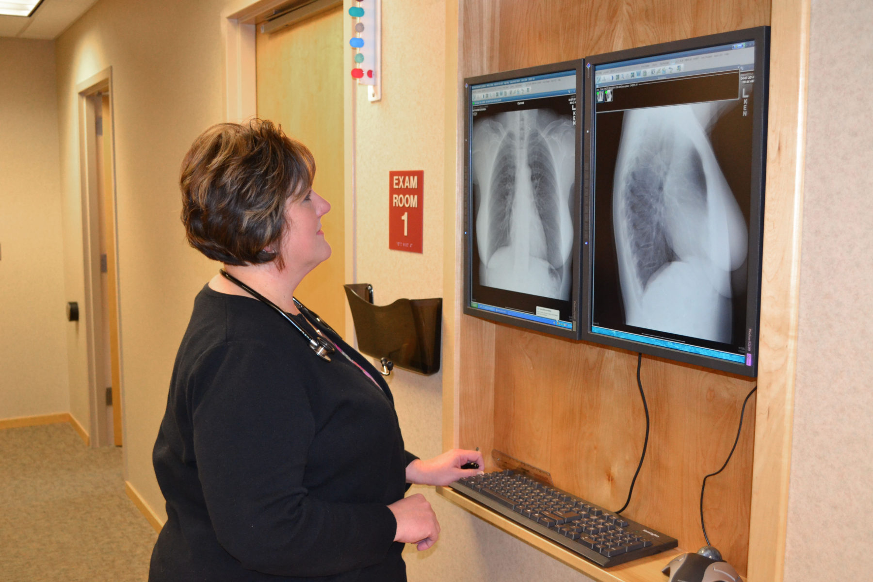
 An X-ray is a quick, painless procedure that takes images of structures inside your body, typically bones and the surrounding soft tissues.
During an exam, X-ray beams pass through your body and are absorbed in different amounts according to the density of the area they pass through.
- How to Prepare
Avoid wearing jewelry or clothing with metal on it, especially if that area is being scanned. You may be asked to undress and wear a gown. Please notify an imaging technologist if there’s a chance you could be pregnant.
- What to Expect
During the exam, a technologist will position your body to obtain the best view of the area being examined. You will be asked to remain still during the procedure to avoid blurring or image distortion. Most X-ray procedures take a 5-15 minutes to complete. - Getting Your Results
A radiologist will read and analyze the images, formulate a report and send the results to your provider. Your provider will contact you to discuss your results.